
Table of Contents
*This post may contain affiliate links. As an Amazon Associate we earn from qualifying purchases.
There are many top ultrasound jobs available today. Discovering which one is right for you is a process. Individuals seeking a career in this field can find information online. The medical practice is continually growing and expanding. There will always be a need for technicians and radiologists employed in this field.
What Is Ultrasound?
Sonography or an ultrasound scan is a procedure used by trained medical professionals that enables the doctor to take a closer look inside the body without making any incisions. Using high-frequency sound waves, it can capture pictures of the interior of the body. This method is preferred as it doesn’t expose the patient or the medical professional to radiation. The pictures that are captured are very helpful in making a diagnosis.
Why Are They Needed?
Soft tissues can be very difficult to see and often don’t show up in x-rays. For this reason, ultrasounds have been used to aid the medical practitioner in discovering a better image of the soft tissue. Ultrasounds are less expensive than other medical procedures making it much more cost effective. This non-invasive procedure is safe and effective.

Ultrasound is safe during pregnancy and therefore, favored by doctors when looking for a non-invasive way to look at a fetus. Because there is not much prep work for the patient other than fasting, it is preferred over other diagnostic methods. The results can be read by a radiologist. A radiologist is a person that has been trained to read the results of the ultrasound.
Why Are They Used?
There are many reasons as to why an ultrasound might be ordered. Usually, an ultrasound is ordered when a doctor desires to see a closer look at something inside the body such as the gallbladder, liver, or heart. If the ultrasound doesn’t produce the desired results, a CT scan or MRI might be ordered next.

CT scans and MRI’s emit radiation and therefore can be hazardous to both the technician and the patient. Medical doctors will consider all options and choose the least invasive option for the procedure at hand. A trained medical doctor will know which option is best suited for the patient.
How Are They Performed?
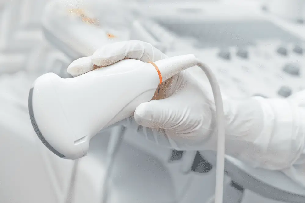
When performing an ultrasound, a technician will use a transducer with a gel-like substance on it. The gel is water-based and safe to use. It helps the transducer roll freely over the skin with minimal friction and aids in helping the sound waves bounce off the body, so clearer results are obtained.

It takes about thirty minutes to an hour to complete the test. While the technician is doing the procedure, they might talk about what they are seeing, but the results will be read in more detail by a medical doctor. A radiologist is qualified to read the results, too.
People working in this profession will prepare the equipment and meet with patients in an exam room. After explaining the procedure to the patient, they will use a transducer to see inside the body and discover any areas that might need further examination. Part of their responsibilities will include helping the patient feel comfortable and informed during the medical procedure.
What Is the Result?
An ultrasound creates a visual image, or a picture of the area viewed. This can be the lungs, heart, liver, or even blood vessels. By taking a picture, it allows the doctor to get a better idea of what is going on in the patient’s body. Sometimes ultrasounds are used to guide a doctor taking a biopsy or performing a procedure inside of the body.

This closer look gives the doctor an inside look at what might be going on in the body. For example, if a patient is having a difficult time breathing, an ultrasound will give a closer look at the heart and arteries. Imagine the benefits of being able to see inside the human body without making any surgical incisions. Ultrasound jobs are beneficial and necessary in the medical field.
Types of Ultrasound
- Transvaginal
- Transrectal
- Transesophageal Echocardiogram
- 4D Ultrasounds
- Echocardiograms
- Doppler
Transvaginal
The transvaginal ultrasound uses a wand called a transducer that is placed inside the vagina to gather images of the surrounding tissue. This non-invasive procedure is a safe way for the doctor to see any areas of concern. It can be used during a gynecological appointment.
Transrectal
Just like the name implies, the transrectal ultrasound is used to take a closer look at tissue inside the rectum. Discovering tumors or cancer cells in advance can help the doctor come up with an effective treatment program. Sonography makes it possible for the doctor to see possible cancerous tissue up close.
Transesophageal Echocardiogram
By placing the transducer inside the mouth and down the esophagus, the doctor can get a better view. This technique allows a doctor to gather safely an ultrasound image that can be read for results. A radiologist can interpret the results seen in the image.
4D Ultrasounds
This is an ultrasound used to get a very clear image. It is most popular with pregnant women seeking a clear image of their unborn babies face. Some results are provided in color now, too.
Echocardiograms
This type of sonogram uses a transducer to examine the chest and heart. A closer image allows the doctor to see any obstructions or things that might impair a patient from a healthy heart. With a clear view of the tissue surrounding the chest, the doctor can make an informed decision and diagnosis regarding the patient.
Bone Sonography
An ultrasound that allows a closer look at bone density is called a bone sonography. It can be very helpful when assessing bone density and possible fractures. A closer look at the skeletal system can help the doctor discover a treatment program for the patient in a non-invasive way.
Doppler
By bouncing high-frequency sound waves, a Doppler is able to show a clear image of what is going on with a patient’s blood flow. This provides a detailed image of a patient’s circulatory system and can show blockages. It is very helpful when viewing an elderly patient. Any time a major surgical incision can be avoided when gathering information is ideal when it comes to aging patients.
